Physical TheraPT

Injury Spotlight

Flexor Hallucis Longus Tendinopathy

IMG_2509.PNG

Often called “dancers tendonitis” due to its prevalence in classical ballet dancers, FHL tendinopathy actually happens to various athletes whose sports require repetitive push-off and extreme plantar flexion (pointing of the foot), such as sprinters, soccer players, gymnasts, and even swimmers.  

The Flexor Hallucis Longus (FHL) muscle is located in the back of the lower leg, originating from the fibula and traveling behind the Achilles tendon. The FHL tendon then passes the inside of the ankle through the tarsal tunnel, and travels along the instep of the foot, ending at the big toe. Its function is to flex the hallux - or big toe. It also has an important role in controlling mid-foot pronation and supination.

Because of these roles, the FHL functions as a powerful convertor of force from the rear foot to the big toe. However, repetitive pushing off the foot and toes can sometimes lead to irritation. This can be worsened when combined with eversion - or an outward motion of the toes relative to the ankle. Often, young dancers will evert when attempting to achieve greater “turnout”, but this also can be seen in runners as excessive pronation - or “arch collapsing” - most often due to strength deficits in the stabilizers of the limb.

medial collapse.jpg

If you’ve irritated this muscle-tendon unit, you may experience pain within the foot or at the back of the ankle depending where along the tendon inflammation has occurred. Some may also experience the big toe “getting stuck” with active movement, or swelling and a crunchy-sensation along the inside of the ankle. Flexing the big toe against resistance, or forcing the foot into a pointed position may also be painful.

While the exact physical cause of FHL injury is under debate, it is believed that the tendon can snag either at the ankle in the tarsal tunnel,  in the mid-foot, or at the sesamoids (two teeny round accessory bones) of the big toe. When combined with repetitive motion, this entrapment of the tendon creates micro-trauma. If left untreated, this can lead to tissue damage. The body's inflammatory response begins to heal these micro-tears, sending more blood and nutrients to the area. This inflammation of the tendon is what is called tendinopathy. 


Restriction of the FHL routinely occurs in three spots:

  • Tarsal Tunnel at inside of ankle, star.

  • Intersection of FHL with neighboring Flexor Digitorum Longus tendon, triangle.

  • Attachment of FHL to the first bone (proximal phalange) of the big toe, square.


To learn more, check out these articles and texts:

  1. https://www.sportsmedtoday.com/fhl-tendinopathy-va-132.htm

  2. Quirk R. Common foot and ankle injuries in dance. Orthop Clin North Am. 1994 Jan;25(1):123-33.

  3. Pagenstert GI, Victor V, Hintermann B. Tendon injuries of the foot and ankle in athletes. Clin Ortho Trauma. 2004; 52(1):11-21.

  4. Simpson M, Howard T. Tendinopathies of the foot and ankle. Am Fam Physician. 2009 Nov 15;80(10):1107-1114.

  5. https://www.sportsinjurybulletin.com/the-flexor-hallucis-longus/#:~:text=Overuse%20conditions%20in%20the%20FHL,posterior%20or%20postero%2Dmedial%20ankle.

  6. Bone Joint Surg. 78A:1491-1500, 1996

  7. Am J Sports Med. 1977;5:84-88

  8. J Orthop Sports Phys 1983; 5: 204-206

  9. Norris R. Common Foot and Ankle Injuries in Dancers. In: Solomon R, Solomon J, Minton S, eds. Preventing Dance Injuries. 2nd ed. Champaign, Ill: Human Kinetics; 2005: 39-51

  10. Foot Ankle Int. 2005; 26: 291-303


Lumbar Disc Pathology

low back pain

Welcome to the club no one wants to be in. Spinal disc injuries are very (very) prevalent, with as many as 20 case per 1000 adults annually. Lumbar disc injuries are the most common, followed by cervical disc injuries. The majority of these cases are injury based, with only 5% being attributed to degenerative disc disease. This month, we’ll explore the anatomy of the spine, break down how disc injuries occur, and share the latest research on recovery.

Back injuries can feel overwhelming. Understanding the structures involved, how they function normally and how they can become dysfunctional, can help focus your rehabilitation goals.

There are 23 discs in the human spine: 6 in the cervical region (neck), 12 in the thoracic region (mid-back), and 5 in the lumbar region (lower back).  Each intervertebral disc (IVD) lies between two adjacent vertebrae in the spinal column, allowing the spine to be flexible without sacrificing a great deal of strength. They also provide a shock-absorbing effect within the spine and prevent the vertebrae from grinding together. 

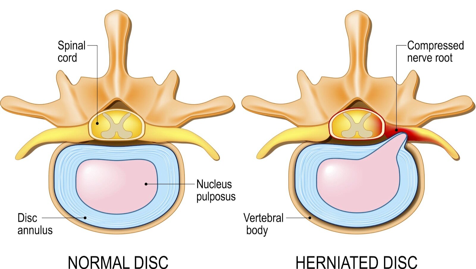
They consist of three major components: the core, nucleus pulposus (NP), the outer ring, annulus fibrosus (AF) and the cartilaginous endplates that anchor the discs to adjacent vertebrae.

So why does it hurt so much, and how does this even happen?!

Disc “bulge”, “protrusion”, “herniation”, even “slipped disc”, as well as disc degeneration all refer to interruption of the normal disc anatomy. Damage to the outer ring, or AF, can occur from sudden trauma to the disc or from disc degeneration due to age and repetitive use. Without the structure of the AF, the NP can be displaced.

While the discs are designed to move to counter spinal movement, repetitive asymmetric compressive loading isn’t ideal. For example, during forward bending, or flexion of the lumbar spine, the NP migrates posteriorly or backwards. Conversely, the nucleus is squeezed anteriorly or forwards during backwards bending, or extension of the lumbar spine. Adding extra weight to one of these positions over and over can cause injury. Research shows the damage to the AF appears to be associated with fully flexing the spine for a repeated or prolonged period of time. 

Like everything else, our Intervertebral discs age. The NP shrinks as it’s gelatinous material becomes dries out over time and is replaced with fibrotic tissue. This places increased strain on the AF. The resulting flattened disc reduces mobility and may impinge on spinal nerves leading to pain and weakness

Due to the proximity of the disc to the spinal cord, if the disc extends beyond its normal resting position, it can result in pain. This pain is due to a combination of the mechanical compression of the adjacent nerve by the bulging NP and localized inflammation and swelling. The symptoms you experience are dictated by what nerves are irritated. Nerve compression can often cause radiculopathy - or radiating symptoms along the path of the compressed nerve into the legs and feet.


To learn more, check out these articles:

  1. Waxenbaum JA, Futterman B. Anatomy, Back, Intervertebral Discs. InStatPearls [Internet] 2018 Dec 13. StatPearls Publishing. Available from: https://www.ncbi.nlm.nih.gov/books/NBK470583/ (last accessed 27.1.2020)

  2. Dulebohn SC, Massa RN, Mesfin FB. Disc Herniation.Available from:https://www.ncbi.nlm.nih.gov/books/NBK441822/ (last accessed 25.1.2020)

  3. L. G. F. Giles, K. P. Singer. The Clinical Anatomy and Management of Back Pain. Butterworth-Heinemann, 2006.

Lumbo-Pelvic Pain in Pregnancy

Whether you’d consider yourself an athlete or not, pregnancy is physically demanding! From start to finish, rapid changes to the female body can greatly impact biomechanics. Rehabilitative medicine and functional conditioning can have a positive impact on the journey.

Radial Nerve Entrapment

Radial nerve entrapment is often thought to be a result of overuse, but it does occur due to other causes - such as direct trauma, fractures, compressive devices, or post-surgical changes. Learn more about how this injury typically presents, and understand the anatomical structures!