Physical TheraPT

Injury & Pain Conditions

Snap, Crackle, Pop

Ever hear a pop when you squat, a crack when you roll your neck, or a grind when you climb stairs? That’s crepitus — the medical term for the noises or sensations that sometimes come from your joints.

Crepitus is common, and in most cases, it’s nothing to worry about. But sometimes, it can signal that your joint mechanics aren’t working as smoothly as they should. Let’s break down why it happens, when it’s normal, and when it may be worth a closer look.


What Exactly Is Crepitus?

Crepitus refers to the popping, cracking, grating, or crunching sounds (or sensations) that occur during joint movement. It can show up in many areas of the body, including the knees, shoulders, hips, spine, and even the jaw.

It’s not a condition by itself, but more of a sign that something is happening in or around the joint. A lot of times, that “something” is completely benign. Other times, it’s a clue that the joint is under stress (Drum et al., 2023).


Why Do Joints Make Noise?

There are several reasons joints produce crepitus, most of which are harmless:

  • Gas bubble formation (cavitation): Joints are lubricated by synovial fluid. When pressure inside the joint changes quickly, gas can form or collapse in the fluid, creating a distinct popping sound (Kawchuk et al., 2015; Fryer et al., 2017).

  • Tendons or ligaments snapping over bone: Soft tissues sometimes shift slightly during movement and then snap back into place, creating a click.

  • Surface changes in cartilage: If the smooth joint surfaces become uneven (such as in arthritis), movement can cause grinding or crackling noises.

  • Internal tissue catching: Soft tissue folds inside the joint, such as plica or meniscal edges, can occasionally cause popping or clicking (Drum et al., 2023).


How Common Is Crepitus?

You’re not alone if your joints make noise — crepitus is widespread, even in healthy people.

  • About 41% of knees in the general population show crepitus.

  • Even among people with no knee pain, roughly one-third still experience it.

  • In individuals with osteoarthritis, the numbers are higher — up to 81% report crepitus (Couch et al., 2025).

The takeaway? Joint noise is very common, and by itself, it doesn’t necessarily mean damage.


When Crepitus Is Harmless

Most of the time, crepitus is simply a mechanical quirk of how your joints move. It’s usually not a concern if:

  • The noise occurs without pain.

  • There’s no swelling, stiffness, or loss of motion.

  • It hasn’t been getting worse over time.

In these cases, there’s no reason to avoid movement — in fact, staying active can keep your joints healthier.


When Crepitus Might Signal a Problem

Crepitus deserves more attention when it’s paired with other symptoms. You may want to consult a physical therapist or physician if you notice:

  • Pain with movement or weight-bearing

  • Swelling, warmth, or stiffness in the joint

  • Locking, catching, or instability

  • Persistent or worsening grinding

  • Loss of function or activity limitations

In some cases, more intense crepitus has been linked with weaker surrounding muscles (like the quadriceps in the knee). This muscle imbalance can place more stress on the joint, making symptoms worse (Jakovacz et al., 2024).

Importantly, while crepitus is more common in people with osteoarthritis, studies show that crepitus alone does not reliably predict disease progression or need for joint replacement (Pazzinatto et al., 2018).


What You Can Do About Crepitus

If your crepitus is painless:

  • Keep moving. Regular activity nourishes cartilage and prevents stiffness.

  • Strengthen muscles. For example, strong quadriceps and hamstrings help reduce stress on the knee.

  • Stretch and mobilize. Increased flexibility can improve tendon and ligament function.


Simple Tools to Keep Your Joints Moving Smoothly

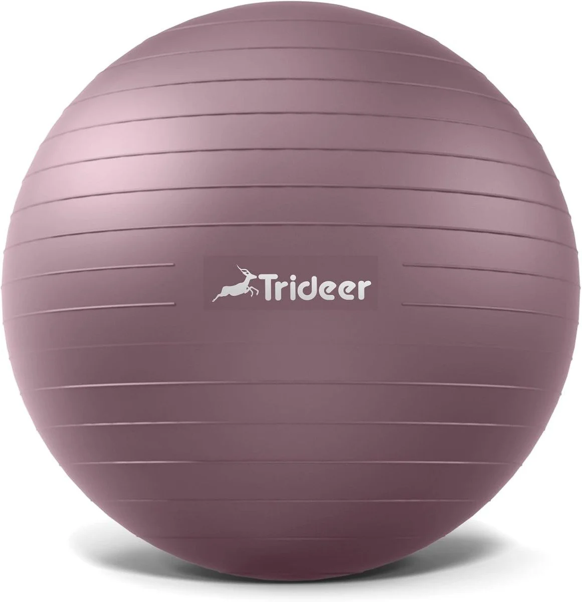
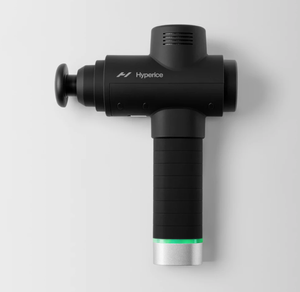
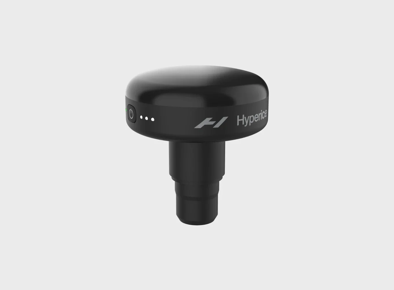
Crepitus can often be eased with tools that promote muscle relaxation and joint mobility. The Hypervolt massage gun with its heated attachment helps improve circulation, reduce muscle tension, and warm up tissues around the joints. Pairing it with gentle movements using an exercise ball encourages controlled mobility and improved joint function.


If your crepitus is painful or comes with swelling or dysfunction:

  • Check in with a physical therapist. They can identify whether the crepitus is harmless or potentially linked to a condition like arthritis, tendon overload, or a fibrocartilage injury (meniscus, labrum).

  • Follow a guided plan. Targeted exercises, joint protection strategies, and activity modifications can help reduce pain and restore confidence in movement.


Bottom Line

Joint noise by itself, even if it sounds dramatic, is usually nothing to worry about. But if crepitus shows up alongside pain, swelling, or loss of function, it’s your body’s way of telling you to pay closer attention.

The good news? Most causes of crepitus respond well to exercise, physical therapy, and healthy movement habits. So the next time your joints crack, remember: noise without pain is normal; noise with pain deserves attention.


References

  • Couch, T. A., Hall, M., Hussain, S. M., Teichtahl, A. J., Wluka, A. E., & Wang, Y. (2025). Knee crepitus: A systematic review of prevalence, incidence, and associations with knee osteoarthritis. Osteoarthritis and Cartilage Open, 7(1), 100471.

  • Drum, E. E., Jauregui, J. J., Probasco, S. K., et al. (2023). Knee crepitus: Current evidence and clinical relevance. Musculoskeletal Care, 21(3), 442–449.

  • Fryer, G., Pearce, A. J., & Herbert, J. J. (2017). Cavitation onset in the metacarpophalangeal joint: A physiological explanation of joint cracking. PLoS ONE, 12(4), e0174190.

  • Jakovacz, N., Mészáros, Z., & Farkas, J. (2024). Relationship between knee crepitus intensity and quadriceps muscle thickness. Manual Therapy, 66, 107102.

  • Kawchuk, G. N., Fryer, J., Jaremko, J. L., Zeng, H., Rowe, L., & Thompson, R. (2015). Real-time visualization of joint cavitation. PLoS ONE, 10(4), e0119470.

  • Pazzinatto, M. F., de Oliveira Silva, D., Azevedo, F. M., & Barton, C. J. (2018). Association between crepitus and progression of knee osteoarthritis: A longitudinal cohort study. Arthritis Care & Research, 70(3), 420–427.

Upper Extremity Sport Durability

For the majority of our upper extremity athletes, the toughest opponent they face is their screen. Here in the Capital of Tech, the hour or two you spend on the field may be the only break from technology. With the challenges of the last year, we’re all spending more time online and on a screen than ever before. While intuitively we all recognize that increased screentime can lead to decreased performance, this post will help break down the impact on upper extremity athletes.

Daily Posture

Start by assessing your resting posture. Look at the position of the head, neck, shoulder and arms relative to the torso. All too often when working on a laptop or handheld device we find ourselves seated with shoulders slumped, looking down, arms and hands together at chest center. Sound familiar?

This will impact your resting breathing posture, as well as your anterior chest and neck muscle tone. The closed position of the throat and rib cage requires the body to work harder and recruit different muscles to inhale and exhale, potentially overusing some muscles in the neck. Adducting the arms, or bringing them together in front of the chest, shortens the muscles of the chest causing them to tighten. The muscles in the back are put in an over-lengthened position, causing them to atrophy due to lack of use. This combination of tight and weak muscles makes it more challenging to find and maintain your athletic posture when you need it.

Finding Balance + Repetitive Behaviors

Using the body described above as our example, the strength imbalance and shoulder position will impact throwing mechanics. To rebalance the system, more emphasis needs to be placed on pulling exercises. This allows the muscles in the upper back to contract while lengthening the muscles in the chest. To compliment this work, incorporate focused dynamic chest stretching and thoracic spine mobility to re-enforce these new postures.

Looking to the back of the body, scapular control is equally essential for throwing mechanics. The scapula has limited ligamentous attachments- it’s the reason the shoulder has such a large range of motion. The catch? The scapula relies more heavily on muscular control than other joints. If you plan on throwing longer and faster, you need to have the eccentric strength to slow all that extra power down. Building a stable foundation (and maintaining it) requires regular rotator cuff strengthening- try incorporating these exercises into your pre-practice warmup or post-strength cool down.

Daily + Weekly Load

Every single athlete, no matter the level, needs recovery days. These low intensity days are the body’s chance to “recharge and download” new movement patterns and muscle fibers. Consistent optimal load to rest ratios will ultimately result in increased durability. To start tracking your load, try this simple low tech strategy:

Upon completing a workout or competition, rate the session using the scale. Multiple that number by the duration of the session in minutes. Track these numbers daily. On a weekly or monthly cycle, aim to create a soft waves rather than dramatic spikes and gaps in activity. Use this chart to plan for important competitions and heavy training days.

 
M.Gibala Ph.D.

M.Gibala Ph.D.


Recovery is not synonymous with rest. Several of our highly competitive athletes have balked at the idea of a rest day, envisioning being confined to the couch for the day. Instead, think about recovery in the following areas:

  • Getting adequate sleep following training (7 hours or more!)

  • Optimizing soft tissues between sessions (massage, foam roll, stretching, etc)

  • Rebalancing consumed macro + micro nutrients (Protein AND electrolytes)

  • Diversifying your skills by trying new sports or activities (VR for reaction time, or swimming for reciprocal rotation)


Summary Block
This block has no content yet. Items you add to the page connected to this block will display here.

Flexor Hallucis Longus Tendinopathy: Part II

FHL treatment

If the symptoms from the last post sound familiar, it’s important to seek care to prevent the condition from advancing. The good news is FHL tendinopathy does not require imaging for diagnosis. You can see a PT or AT directly- they’ll evaluate and diagnose your condition, and will refer you to a physician as needed.

After assessing your leg, your clinician will be able to ascertain contributing factors that led to the injury and how to address them with an individualized exercise program including neuromuscular re-education and functional training. Neuromuscular re-education helps reprogram the way you move- focusing on specific muscle activation and timing. This strategy takes the whole kinetic chain into consideration, looking for inefficient habits that may seem unrelated to your injury.

For example, many athletes diagnosed with FHL tendinopathy present with excessive pronation- or arch collapsing- a faulty pattern that can contribute to overloading the FHL in running. Strengthening the hip, pelvis and core muscles, combined with improved running mechanics can reduce this problem for the long term!

If you are able to see a clinician in-person, manual therapy may be beneficial to reduce inflammation, muscular tension, and any joint stiffness.  

Until your appointment, follow these recommendations at home:

  1. Rest from the aggravating activity - running, dancing, etc.

  2. For pain management, try over the counter NSAIDs (ie Advil, Aleve)

  3. Use contrast therapy to reduce inflammation:

    • Submerge foot for 10-15’ total following the pattern below:

      • 2-3’ in ice water

      • 2-3’ warm water

      • 2-3’ ice water

      • 2-3’ warm water

      • 2-3’ ice water

  4. Self-massage the outer calf by rolling with a tennis ball or something similar on the floor

  5. Perform a seated calf stretch with a towel at the toes, being sure to pull the big toe back with the foot

Flexor Hallucis Longus Tendinopathy

IMG_2509.PNG

Often called “dancers tendonitis” due to its prevalence in classical ballet dancers, FHL tendinopathy actually happens to various athletes whose sports require repetitive push-off and extreme plantar flexion (pointing of the foot), such as sprinters, soccer players, gymnasts, and even swimmers.  

The Flexor Hallucis Longus (FHL) muscle is located in the back of the lower leg, originating from the fibula and traveling behind the Achilles tendon. The FHL tendon then passes the inside of the ankle through the tarsal tunnel, and travels along the instep of the foot, ending at the big toe. Its function is to flex the hallux - or big toe. It also has an important role in controlling mid-foot pronation and supination.

Because of these roles, the FHL functions as a powerful convertor of force from the rear foot to the big toe. However, repetitive pushing off the foot and toes can sometimes lead to irritation. This can be worsened when combined with eversion - or an outward motion of the toes relative to the ankle. Often, young dancers will evert when attempting to achieve greater “turnout”, but this also can be seen in runners as excessive pronation - or “arch collapsing” - most often due to strength deficits in the stabilizers of the limb.

medial collapse.jpg

If you’ve irritated this muscle-tendon unit, you may experience pain within the foot or at the back of the ankle depending where along the tendon inflammation has occurred. Some may also experience the big toe “getting stuck” with active movement, or swelling and a crunchy-sensation along the inside of the ankle. Flexing the big toe against resistance, or forcing the foot into a pointed position may also be painful.

While the exact physical cause of FHL injury is under debate, it is believed that the tendon can snag either at the ankle in the tarsal tunnel,  in the mid-foot, or at the sesamoids (two teeny round accessory bones) of the big toe. When combined with repetitive motion, this entrapment of the tendon creates micro-trauma. If left untreated, this can lead to tissue damage. The body's inflammatory response begins to heal these micro-tears, sending more blood and nutrients to the area. This inflammation of the tendon is what is called tendinopathy. 


Restriction of the FHL routinely occurs in three spots:

  • Tarsal Tunnel at inside of ankle, star.

  • Intersection of FHL with neighboring Flexor Digitorum Longus tendon, triangle.

  • Attachment of FHL to the first bone (proximal phalange) of the big toe, square.


To learn more, check out these articles and texts:

  1. https://www.sportsmedtoday.com/fhl-tendinopathy-va-132.htm

  2. Quirk R. Common foot and ankle injuries in dance. Orthop Clin North Am. 1994 Jan;25(1):123-33.

  3. Pagenstert GI, Victor V, Hintermann B. Tendon injuries of the foot and ankle in athletes. Clin Ortho Trauma. 2004; 52(1):11-21.

  4. Simpson M, Howard T. Tendinopathies of the foot and ankle. Am Fam Physician. 2009 Nov 15;80(10):1107-1114.

  5. https://www.sportsinjurybulletin.com/the-flexor-hallucis-longus/#:~:text=Overuse%20conditions%20in%20the%20FHL,posterior%20or%20postero%2Dmedial%20ankle.

  6. Bone Joint Surg. 78A:1491-1500, 1996

  7. Am J Sports Med. 1977;5:84-88

  8. J Orthop Sports Phys 1983; 5: 204-206

  9. Norris R. Common Foot and Ankle Injuries in Dancers. In: Solomon R, Solomon J, Minton S, eds. Preventing Dance Injuries. 2nd ed. Champaign, Ill: Human Kinetics; 2005: 39-51

  10. Foot Ankle Int. 2005; 26: 291-303


Back At It

OverPage-0 41.png

To wrap up our section on lumbar disc injury, Nina and I break down some of the most common patient questions we receive. As an AT, Nina is accustom to triaging athletes in pain, helping them find confidence in their plan of care.


SV: For the past month, we’ve been sharing the latest research on lumbar disc pathologies. As a recap, what are your top three take aways for experiencing back pain?

NS: We’ve covered a ton of information on this topic recently, looking at the impact of pregnancy as well as injury. The overall takeaway is that each individual’s experience can be different- this goes for symptoms and solutions. That’s why personalized care can have such a big impact for back pain. Similarly, there isn’t one guaranteed strategy for managing a lumbar disc injury. A combination of treatments, including soft tissue work, stability and mobility exercises, and pain management, is typically needed. Last, use this as an opportunity. For most people, an injury like a disc herniation can be a catalyst to make adjustments to their behaviors, allowing them to emerge stronger than before.

SV:  How does this differ from the type of back pain caused by pulling a muscle?

NS: Discogenic pain has a different quality; many patients report feeling sharp, electric sensations, while others complain of weakness. Nerve pain can “travel,” sending irritation down one or both legs. Muscle spasm often accompanies a disc injury- it’s a protective strategy. Muscles of the back will automatically tighten above and below the injured disc to limit movement. This tightening makes it difficult to use muscle systems, or groups of muscles designed to support compound movements, effectively.

Pulling a muscle, or overusing a muscle, can usually be correlated with a specific action or activity. You may feel tight, or restricted. This should resolve, or feel considerably better, in 72 hours if it’s just muscular.

SV: Here’s a popular combo question- do patients need to get imaging for this type of injury? How likely will they need surgery?

NS: It’s a valid question- too often, we only hear about worst case scenarios for injuries. Unless you’ve suffered a traumatic injury, like a car crash, we don’t typically recommend imaging right away. Rehab exercises and pain management will be the most productive first steps. If you eventually need surgery down the road, being as strong as possible going in will dramatically help your recovery!

Surgery makes the most sense for patients who have a high risk of complications- like if your herniation or displacement is so severe you may cause permanent damage to your spinal column- or for those who haven’t succeeded with conservative care after a number of months.

We’re big believers that patients should be the center of their plan of care, with a variety of clinicians and specialists, coaches and supporters surrounding them. Without a different perspectives, you can overlook options. Ask questions, get second options- be your best advocate!

SV: Once someone injures their back, does that mean they can never workout or play sports again?

NS: In the vast majority of cases, definitely not! Most patients are able to return fully to their previous level of activity. Like any major injury, it will take dedication and focus to recover. It’s essential to allow your body enough time to heal, and to correctly learn, or re-learn, movement patterns. Look for rehab practitioners that understand the demands of your activity, and can help get you there. Stopping at 65-75% better increases your chance of re-injury significantly.

SV: With back pain being one of the most prevalent injuries, how can someone lower their risk for injury?

NS: Regular physical activity will help to protect against a number of ailments and injuries. Particularly with the recent transition to virtual learning and working, movement is even more important, as we are naturally more sedentary in this scenario. Adding in tri-planar exercise, including linear, lateral and rotational work, can help make sure you’re prepared for whatever life throws you! If you’re new to exercise, or struggling with home exercise routines, setting up an appointment to work with a clinician or coach can give you the confidence you need to move safely.

SV: At Physical TheraPT, as many of our patients know, we love staying current on the latest innovations in exercise and rehab equipment. What’s your favorite product out there right now for back pain patients?

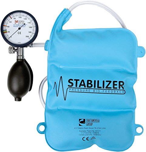
NS: For patients still in the early stages of recovery, I really like the biofeedback cuff. This deconstructed blood pressure cuff helps increase awareness of pelvic stability and highlights how well someone is able to maintain a braced position when you add on movement. For those who are out of pain, and show progress with more challenging exercises, the 3D strap is my favorite. Don’t be deceived by the simple nature of this piece of equipment! It adds rotational load to almost any movement pattern, allowing athletes to adapt and practice in a safe environment.

Screen Shot 2020-10-12 at 11.54.39 AM.png

SV: Last question- what’s your go-to strategy for pain relief with this type of injury?

NS: Finding a position of comfort that you can rest in. Back injuries can feel unrelenting- having a position that gives you some relief is critical in the beginning stages of recovery. Laying on a firm surface, resting on your back with knees bent and feet planted, referred to as Hook-Lying Position, is generally safe for most patients. Others may find relief with a bit of light traction, leaning into your elbows at a counter, or floating in a pool.